Teaming up for good science!
Being a team player is not only important in sports. In science, too, researchers with different skill sets and backgrounds work as a team towards a common goal. In research, the term “team science” refers to the collaborative effort to address a scientific question, often involving teams from different fields and geographical locations.
As an example of team science, here, we are going to tell the story of three research groups studying the brain in different countries across Europe and how they came together to collaborate on the same project.
In Barcelona, a neurosurgical research team observes that electric stimulation of a specific area of the human brain provokes language errors during awake brain surgery. When the surgeon pokes that brain area with the stimulator while showing pictures to the patient, they call an elephant a zebra, or an apple, a pear.
Meanwhile, in the Dutch town of Nijmegen, another team of researchers explores the pattern of brain activity which may allow people who have suffered a stroke to recover their language skills.
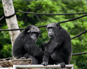
The third team, based both in Nijmegen and Oxford, explores whether the anatomy of a chimpanzee’s brain is similar to that of a human using structural brain imaging.
It might sound like the work of the three groups is unrelated to each other, but the truth is that the three teams were centred on one specific part of the brain – a crucial region for human language located in the temporal lobe.
To better understand this part of the brain, the three teams decided to join forces and collaborate in a multidisciplinary study. But how did the idea of a collaboration start?
The paths of the three teams crossed in Nijmegen. It is becoming more and more common for researchers to travel to other countries to do placements for a few months or carry out their research there for a few years. This is often called research mobility and gives researchers the opportunity of learning new skills and being exposed to different research cultures.
When Joanna Sierpowska finished her PhD in Barcelona, she moved to Nijmegen to do a postdoc in the group of Vitória Piai at the Donders Institute. Thanks to the scientific exchange offered by this centre through regular meetings and conferences, an interesting poster caught Sierpowska’s and Piai’s attention. The poster, presented by Katherine Bryant from the group based between Nijmegen and Oxford, showed white matter tracts in the chimpanzee’s brain. After a lively discussion between the three of them and the head of the Cognitive Neuroecology Lab, Rogier Mars, the idea of a collaboration started.
The researchers agreed that they would study the pattern of white matter connectivity of crucial portions of the temporal lobe with the rest of the brain, comparing humans and chimpanzees. And here is where the second benefit of teaming up for science becomes aparent. New research idea could be explored straight away by working on already existing datasets. The chimpanzees’ data was made available by the the US-based National Chimpanzee Brain Resource (chimpanzeebrain.org). Data from healthy participants was shared by one of the researchers working on this study, Nikki Jansen, who acquired the data for her own anatomical inquiries.
Sharing and opening researchers’ datasets is becoming the norm. It helps to not only make the research process transparent and trustworthy, but also to facilitate research and avoid unnecessary, costly acquisitions (which is in line with the concept of “green science”). Needless to say, it also fosters building a collaborative research network worldwide.
Going back to this multicentric study, what were the researchers looking for? At first glance, the brains of humans and chimpanzees look very much alike. Yet, we know that there are some striking differences between the two species. One of the most obvious differences is the unique ability to communicate through language that humans have.
Understanding what characteristic of the human brain could have enabled language throughout evolution has inspired many studies. However, most of them have focused on a white matter tract that connects the temporal and frontal lobes known for being involved in language: the arcuate fasciculus. The multidisciplinary team was interested in a distinct approach.
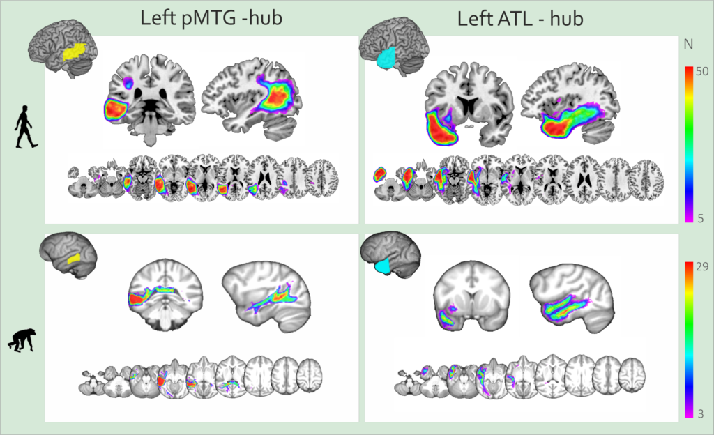
They wanted to study the connectivity of the anterior and posterior areas of the temporal lobe. The choice of these regions of interest was not random. They are two major hubs related to language. The posterior middle temporal gyrus (pMTG) is involved in assigning meaning to words and processing syntax. A lesion in this area can impair the ability to understand the appropriate meaning of a word depending on the context. On the other hand, the anterior temporal lobe (ATL) is involved in forming representations of the meaning of words. Patients with semantic dementia, a neurodegenerative disease affecting the ATL, show problems remembering general concepts about objects and events, as well as the words used to describe them.
In addition to the existing scientific literature about the pMTG and the ATL, the team relied on their own clinical experience. Sierpowska and Piai observed patients undergoing brain stimulation during surgery and patients recovering from stroke. In both cases, these temporal brain regions had a key role in language. If they were damaged or lost, the affected person struggled with speaking. For example, they could have problems finding the appropriate words to express their thoughts. That made the researchers think that there could be something unique in humans in how these areas are connected to the rest of the brain.
To investigate the white matter fibres connecting these two hubs with the rest of the brain, the teams used a neuroimaging modality called diffusion-weighted imaging (DWI) to scan the brains of 50 humans and 29 chimpanzees. This neuroimaging modality allows researchers to effectively (and beautifully) show the anatomical white matter fibres connecting the distinct portions of the brain.
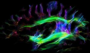
The visualization of white matter tracts at the level of arcuate fasciculus (c) A. Anwander, MPI CBS
When comparing the two species, the researchers found that the posterior temporal areas were mainly connected to other areas of the temporal lobe. However, in humans, new connections emerged. The human pMTG showed more connections with frontal and parietal lobes, using the arcuate fasciculus as an anatomical avenue, while the human ATL showed more robust connections with ventral white matter tracts.
These findings, although purely anatomical, shed light on the evolutionary changes underlying human language. The fact that this pattern of connections is so unique to humans, together with the clinical data, suggests that these new connections may have enabled our full-fledged language abilities.
In addition, the results of this study could help guide clinical interventions as well as clinical neuroscience research questions.
Lastly, this research shows the importance of team science and multidisciplinary collaboration, as the study would not have been possible without all of the researchers involved.
Authors: Clara García Gorro & Joanna Sierpowska
To find out more, check the original research published in PNAS: https://doi.org/10.1073/pnas.2118295119
This article received funding from ”la Caixa” Foundation (ID 100010434) and from the European Union’s Horizon 2020 research and innovation programme under the Marie Skłodowska-Curie grant agreement No 847648/ LCF/BQ/PI21/11830014 awarded to Joanna Sierpowska.
No Comments